Thursday, March 17, 2016

The Mammoth in the Room


Last year, an extremely well-preserved carcass of a dog from the Pleistocene era (12,400 years ago) was unearthed from its icy grave in Tumat, Russia.  Among many present for its recent autopsy, was Hwang Woo-Suk, a South Korean professor who hopes to use some of samples he collected to bring the extinct animal and others such as the woolly mammoth back to life through de-extinction.
               De-extinction can be defined either as the rebirth of an animal that was extinct or the creation of an animal that greatly resembles an extinct species.  Several possible techniques exist for “resurrecting” these one thought lost animals.  One of the most promising methods is use of CRISPR/Cas9 to “cut and paste” genes from an extinct animal into those of a living common ancestor.  This method is being used by George Church’s lab at Harvard University to replace 14 loci of an elephant genome with a mammoth version of those sequences.   Currently, they are only working on transforming these cells into tissues and stem cells but it is estimated that within a decade, it could be possible to meet a breathing woolly mammoth.  Another possible method for de-extinction is cloning, where DNA from a recently extinct animal is inserted into the egg another animal, then implanted into a surrogate mother.  This method was used in 2009 to resurrect the bucardo, an ibex species that went extinct in 2000. The clone, however, died just minutes after its birth from deformities.   Due to the severity of clone deformities and shortened lifespans due to telomere shortening, it is likely to be a while before cloning can be used in this method with success.
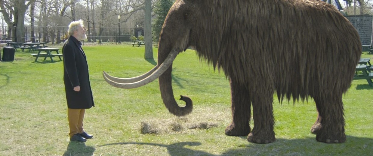
               With scientists closing in on reversing extinction, it is easy to imagine a real-world Jurassic Park filled with extinct animals like the Woolly Mammoth, Passenger Pigeon, Dodo, and Tasmanian Tiger.  However, also like in Jurassic Park, grave endings may result from “playing God”.  When re-introduced into the wild, the revived creatures may turn out to be invasive species in an ecosystem that, after thousands of years, has moved along without them and assumedly achieved equilibrium in their absence. Another worry is that if extinction is no longer a finality, conservation efforts may cease.  This leaves many wondering if it is better to spend money on the revival of a species no longer suited to survive or on endangered species.  

Boyle, Jamie. "Science Explainer: We May Not Resurrect Dinosaurs but Other Extinct Animals Likely to Be Revived | Genetic Literacy Project." Genetic Literacy Project. The Genetic Literacy Project, 16 July 2015. Web. 15 Mar. 2016.
Choi, Charles. "First Extinct-Animal Clone Created." National Geographic. National Geographic Society, 10 Feb. 2009. Web. 15 Mar. 2016.
Liesowska, Anna. "Ancient Puppy's Brain Is 'well Preserved'... as Dog Bares Its Teeth after 12,400 Years." RSS. Siberian Times, 16 Mar. 2016. Web. 16 Mar. 2016.


Monkeys Move Wheelchairs With Their Minds

First off, no, monkeys have not developed telekinetic powers and then attempted to use them against the disabled. That I know of.  The involvement of monkeys in scientific research seems to somehow generate absurd situations that leave the casual observer bewildered and dubious. This is most certainly the case for a recent study that sounds like something straight out of a futuristic sci-fi novel. 
Researchers at Duke Health have recently created a brain-machine interface (BMI) that allows primates to move a robotic wheelchair using only their thoughts. The experiments began in 2012, when hundreds of microfilaments were implanted in the premotor and somatosensory regions of the brains of two rhesus monkeys.  The monkeys were trained by passively moving the chair to the target, a bowl of grapes, while the researchers recorded the large-scale electrical brain activity. The motions of the wheelchair where correlated with signals from different neurons in the monkeys’ brains, creating computational algorithms that mapped brain activity to different trajectories.
An overview of the experimental design
The data from this initial experiment was then used to create the interface system, which detects signals in the monkeys’ brains, and then translates them into commands that move the wheelchair.  The monkeys’ skills at moving the chair actually improved as the experiment continued, with one of the monkeys going from completing the task in 43.1 seconds to 27.3 seconds.  Other differences in brain activity were noticed when the monkeys themselves were moving the chair, as opposed to when they were merely passengers, and their neurons became better attuned to the distance between the chair and the grape dispenser.  This shows promising evidence that the brain is able to adapt to and assimilate the device. According to researchers, the next step in development will be to record more neuronal signals in order to increase the accuracy and fidelity in the primate model before beginning any trials with the device implanted in humans.
A monkey successfully maneuvers the chair to the dish of grapes
This is more than just simple monkeyshines. For many individuals with disabilities that impair movement, wheelchairs are still the primary mode of transportation.  Currently, there is a large amount of focus on the development of exoskeletons controlled by signals from external electroencephalography (EEG), but these devices have been developed for paraplegic patients.  ALS patients or those who are quadriplegic would most likely not be able to use these devices, but removing the need for someone else to move their wheelchair could give them more independence, autonomy, and dignity.  There is even evidence that BMIs can lead to partial neurological recovery by triggering cortical plasticity.  Though there is still much more work to be done to increase the accuracy of intracranial interfaces, it is clear that BMIs could be of great help to the physically disabled.

 Sources:

Davis, Nicola. "Monkeys Taught to Control Robotic Wheelchair by Thought Alone." The Guardian. Guardian News and Media, 03 Mar. 2016. Web. 17 Mar. 2016.

Rajangam, S. et al. Wireless Cortical Brain-Machine Interface for Whole-Body Navigation in Primates. Sci. Rep. 6, 22170; doi: 10.1038/srep22170 (2016).

Wednesday, March 16, 2016

Inking the Immune System?

Many people assume that tattoos are bad for your body, injecting a foreign object into your body and attempting to make it permanent.  People are scared of the cleanliness of the needles, what rashes, and infections they may get from the tattoo itself.  Actually, in some cases this assumption is correct.  A small percentage of people do get allergic reactions or skin injections after they get a tattoo.  However, a study was done on people who had gotten tattoos.

In the study, they took saliva samples before their tattoo and afterwards.  This study showed that directly after getting their first tattoo, people’s immune system lowers temporarily.  The immune system uses up antibodies fighting possible infection at that tattoo site.  Through the saliva samples, the researchers tested the levels of immunoglobulin A.  They found that the levels dropped significantly after getting their first time.  In people who got tattoos more often, this lose in immunoglobulin A was much less.  The immune system works so hard to try to fight off the foreign substance being put into your body, that it’s focused on that and only that which can cause you to feel tired, exhausted, and it’s easier to get sick.

However, the more tattoos you get, the stronger your immune system will become.  Picture getting more tattoos like training for a marathon.  At first you feel really out of shape, but after weeks of training you’re stronger than you’ve ever been and know you can run a marathon.  This is because after the tattoo, your body returns to normal.  Moreover, if you continue to get more tattoos, your body will feel that stress but won’t return to that same set point.  Instead, the body will adjust that set stress point to a higher level which makes your immune system stronger.

So, if you have a relative or a friend who constantly disproves tattoos make sure you tell them that tattoos are beneficial as well.  You can show them the studies that these scientists have done to prove that many tattoos benefit the human body and the immune system itself.


Goodbye Shuteye?

Sleep, at least for me, is definitely a novelty since starting college. Even in high school, I kept myself very busy and often traded in a good night’s sleep for homework, soccer practice, or my job. I knew that I should probably be getting more sleep, but how much? And how important is it to get the suggested amount? The National Sleep Foundation recommends that teens (14-17) get 8-10 hours of sleep each night, while adults (18-64) only require 7-9 hours.  However, a 2009 study conducted by the NSF predicted that the average American only gets 6.7 hours of sleep during the week.


As you can see, the average amount of time people have been sleeping each night has been steadily declining. Dr. Raj Kakar, a medical director at the Dallas Center for Sleep Disorders, believes that stress is the main culprit of sleep loss. His findings correlate with the American Psychological Association’s survey in which 52% of respondents (out of 7,000) reported losing sleep at night due to stress.  Stress isn’t the only culprit of sleep loss; day-to-day distractions also play a role in sleep loss. With the advancement of technology, distractions like cell phones, tablets, and other devices keep people awake and busy longer than before. The blue light emitted from devices can prevent the pineal gland from releasing melatonin, a hormone associated with sleep onset. Therefore, even when people finally get to bed, it is harder to fall asleep.

The big question is, how much does lack of sleep affect our daily lives – and possibly long-term health? The National Highway Traffic Safety Administration estimates that fatigue is a cause in 100,000 car crashes in the U.S., with the problem being greatest among people under 25 years old. Additionally, sleep plays a large role in critical thinking and learning. Therefore, a lack of sleep has shown to impair alertness, concentration, reasoning, and problem solving. Shockingly, sleep loss can be linked to weight gain. A peptide hormone called ghrelin, also known as the “Hunger Hormone”, stimulates hunger, while leptin, the “Satiety Hormone”, inhibits hunger. Shortened sleep time has been associated with an increase in ghrelin and a decrease in leptin. Finally, sleep loss can have an effect on long-term health, as it can be quite harmful to the brain. An interesting graphic to the right shows a variety of harmful side effects of sleep loss, perhaps the most worrisome being brain damage as a result of pulling too many all-nighters.


Get some sleep! Power down, calm down, and lie down!


Works Cited
1. Fisher, Theresa. "What Sleep Deprivation Does to Your Brain, in One Stunning Infographic." Mic. 18 Nov. 2014. Web. 16 Mar. 2016.

2. "NSF Recommends New Sleep Times." National Sleep Foundation. 2 Feb. 2015. Web. 15 Mar. 2016.


3. Park, Madison. "Why We're Sleeping Less." CNN. Cable News Network, 6 Mar. 2009. Web. 15 Mar. 2016.

"Person on a Chip"

Being placed on an organ transplant list is a nightmare for patients that are racing the clock to prevent premature death. Each second ticking by is another moment of terror for them. But the fear of finding hearts, livers, and, potentially, other organs in time may be diminished in a few years. Around the world research groups, as well as a team from University of Toronto, are trying to find ways to grow tissues under lab conditions that “mimic a real person’s body” (Mole, 2016). At the University of Toronto, chemical engineer Milica Radisic, graduate student Boyang Zhang and the rest of their research group have created a method in growing these tissues.

The team has created the AngioChip, “a fully three-dimensional structure complete” with a vascular system. The scaffold is made out of a polymer that is both biocompatible and biodegradable. The polymer is then used, in thin layers, to create the structure of the AngioChip. The layers, resembling a computer chip (hence the name), are stacked into a three-dimensional structure of synthetic blood vessels by using UV light to cross-link the polymer and bond it to the previous layer. The finished structure is submerged in a liquid containing living cells of the organ they want to build. These cells then attach to the inside and outside channels of the scaffold and begin growing (Irving, 2016).

A close-up of the AngioChip.
Using this method, the team had built model version of both the liver and heart tissues that are functionally similar to the real thing. The model liver “actually produced urea and metabolized drugs.” The group has also injected white blood cells into the connected heart and liver apparatus and observed that the blood cells moved, as they would have in a human. Other than the transplant possibilities for the models, they have great potential in the field of pharmaceutical testing. Instead of animal testing and controlled clinical trials, the lab-grown human tissues would provide a realistic model to test drugs. The tissues can also be used to “validate the effectiveness of current drugs” and screen a lot of chemical compounds to discover new drugs (Univ. of Toronto, 2016).

When seeded with heart cells, the flexible polymer scaffold contracts with a regular rhythm, just like real heart tissue. Credit: Boyang Zhang
Even though this is a huge advancement in medical technology, there are still many hurdles to overcome. Each AngioChip is made by hand; if the chip is to be used industrially, a thought-out machine manufacturing process needs to be created to produce mass quantities in a short period of time (Univ. of Toronto, 2016).


As a chemical engineering student, I am intrigued about the applications chemical engineering has in the medical field. I desire to pursue a career that has a focus on medicine and this article displayed a use chemical engineering has in creating a potentially life saving device. I am very excited about this tissue engineering advancement since the same process can be used to create other major vascular organs of the human body. This will immensely benefit the patients that are waiting for an organ donor match, which is the major drawback of organ donation. However, the tissue cultures for the AngioChip can be engineered to be genetically identical to the intended host, reducing the risk of organ rejection (Mole, 2016). While there is still manufacturing kinks to work out, the huge benefits that can come from this invention will revitalize organ donation. However, there may be social consequences to this: will people be more likely to practice bad habits, such as smoking and excessive alcohol intake, since there will be a viable organ essentially waiting for them?

References:

Irving, Tyler. "'Person-on-a-chip' - U of T Engineers Create Lab-grown Heart and Liver Tissue for Drug Testing and More - U of T Engineering News." U of T Engineering News. 07 Mar. 2016. Web.

Mole, Beth. "Creation of Mini-organs Follows Mini-brains." Arstechnica. N.p., 9 Mar. 2016. Web.

University of Toronto Faculty of Applied Science & Engineering. "'Person-on-a-chip': Engineers grow 3-D heart, liver tissues for better drug testing." ScienceDaily. ScienceDaily, 7 March 2016.

Monday, March 14, 2016

Making the Lame Walk

     There is hope for quadriplegic and paraplegic people and animals in the future.  In April of 2013, the University of Washington and the Japan Science and Technology Agency in Tokyo, Japan published an article that stated work on using artificial corticospinal and musclospinal connections to restore pathways in the spine had been successful.  The testing was performed on a monkey with a C-2 injury causing paralysis from the shoulders down.

      As a bit of background information, a spinal cord’s damage creates a lesion in the spinal network.  The nerves above and below the lesion are still functional, but because of the break in the path, the nerve signals being sent below the lesion are unable to reach the brain and cause movement.  This is why paralysis is a result of spinal cord injuries.  With the research at Washington University, a “neural bridge” could connect the two nervous pathways and could possibly restore functionality.

     The testing for this experiment is as followed.  The monkey with the cervical spinal injury was anesthetized and cortical implants were placed in the dura, finger, wrist, arm areas that were considered the primary motor cortex, and the dorsal arm areas for premotor cortex close to the lesion in the C-2 vertebrae. 

     For three months after the lesion, the monkey was unable to move its fingers independently at the level that it was pre-lesion.  After the 5-7 week mark, the ability for a “power grip” gradually came back but not at a promising level.  This result reflected another study that was done two years earlier.  Mapping of the electrode sights was completed throughout the trial and it was found that 40% of the electrodes could provoke movement.  The loss of function in the upper extremity was consistent with another study that was performed in 2004.

     It’s obvious that the research done hasn’t accomplished a total solution to “curing” paralysis, but with more time, money, and brain power solutions could be produced to get the other 60% of the electrodes working.  If this study were to produce a way to bridge the gap between the superior and inferior nerve ends of a lesion then the benefits would certainly outweigh the time and multiple experiments it took to achieve such a monumental discovery.

Citations: 
Nishimura, Yukio, Steve I. Perlmutter, and Eberhard E. Fetz. "Restoration of Upper Limb Movement via Artificial Corticospinal and Musculospinal Connections in a Monkey with Spinal Cord Injury." Frontiers. Frontiers Media S.A., 11 Apr. 2013. Web. 14 Mar. 2016.

Sunday, March 13, 2016

Ebola: Africa's Ecological Guerrilla Fighter


Guerrilla warfare usually refers to the strategy of fighting by groups of irregular troops within areas occupied by the enemy, using tactics such as surprise, ambush, and deception (Smith 1). A small group of commandos will remain in hiding until the correct moment to raid the enemy, performing a quick strike, and then retreating to the underbrush where they are concealed from view. This action is repeated as long as it is necessary, until a significant amount of damage has been done. Guerrilla warfare has been a very successful attack design for a wide array of people and organisms, including a virus that has recently become one of the most feared diseases among the United States population. The Ebola virus’ has utilized this tactic, which is the main reason it has been so efficient in human lethality and individual survival.

Ebola is a zoonotic pathogen; it is able to cross from an animal host hideaway into a human, spreading uncontrollably at times and leading to death. This type of virus enables Ebola to have a very complicated and problematic advantage: the ability to hide. It is not hiding consciously of course, but in a place it is able to survive and reproduce. This area of hiding is primarily an organism, called a reservoir host, which carries the pathogen but has little to no suffering or illness. Ebola is able to lurk in an ecosystem where there is much diversity and the environment remains relatively undisturbed. But once an ecological disturbance has occurred, Ebola emerges from depths of the jungle. It makes a quick strike, infecting then killing numerous people and then disappearing again into the unknown. This happens continuously, using a form of guerrilla warfare, teeming with surprise attacks to take human and animal lives before retreating to its reservoir host.

Because of Ebola’s method of irregular and ambush infections, it has proven difficult to study the virus. So far no proven treatment or cure has been identified, which has created a very high mortality rate for the disease. Ebola is transmitted through bodily fluids, so infection can spread rapidly as a sudden surge of sickness; people will not know something is contaminated until it is too late. As of now, the identity of Ebola’s reservoir host remains unknown, although some recent evidence has pointed to a type of fruit bat located in the caves of West Africa. As long as scientists cannot pinpoint the exact host, Ebola will continue to inflict its terror on humans in West Africa and the rest of the world.

Currently there are at least five known strains of the ebolavirus. Four are scattered across sub-Saharan Africa and one seems to be endemic to the Philippines and has traveled to the United States on occasion. These different forms have various symptoms, and each new virus discovered seems to be more lethal, as it increases its death rate progressively. This means that the Ebola has been evolving and continues to evolve, making genetic changes that favors its survival and increases its destructiveness. This fact creates an eerie sense that Ebola is not gone for good; its reappearance is inevitable. It is scary to think that the virus is becoming worse, and eventually an outbreak will occur where it spreads even faster and causes more casualties. Maybe the next occasion Ebola happens to emerge from its quiet hideaway, it will not be contained in just Africa and will spread to other countries more than it has previously. With no known remedy, this could lead to the worst outbreak the world has seen in years.


References:                                                                                                                                

Quammen, David, and David Quammen. Ebola: The Natural and Human History of a Deadly Virus. N.p.:      n.p., n.d. Print

Smith, Peter. "Guerrilla Warfare." TheFreeDictionary.com. N.p., n.d. Web. 13 Mar. 2016.

Genetic Testing and its Ambiguity


                Continuing the discussion from last week of genetic testing, this article is based upon Angie Watts, a 44 year old woman suffering from breast cancer. Unlike Huntington’s Disease, which is a dominate condition meaning if you possess the gene, there is a one hundred percent chance that you will develop the disease, the genes linked to breast cancer do not give definitive answers. Even with the $215 million dollar precision medicine initiative from President Obama, genetic testing and genetically personalized medicines from cancer are proliferating, but unfortunately not in the case of breast cancer. Despite the genetic data now available, doctors have not been able to decipher the clues behind this disease.
                Angie Watts received a lumpectomy for her breast cancer last year and was ready to start her radiation therapy, yet her doctor shocked her with the news that she had inherited a specific gene alteration that may induce the growth of her cancer if radiation was applied. Due to this issue, her doctor suggested a double mastectomy. The next doctor she spoke to advised her to go with the radiation therapy as he claimed the mutation in her genes was not known to be harmful. Going from doctor to doctor, Ms. Watts heard new sides of the story until a group of doctors finally came together and told Ms. Watts that the decision was up to her.
                The story of Ms. Watts shows how much more is left to learn about genetic testing and how it can be applied to cancers, especially breast cancer. Although we have all of this technology and can sequence genes relatively easily, we don’t necessarily know what the genes and mutations mean yet. The main issue comes when there are multiple mutations found in a person’s genome. Many of these mutations will come together and it can be extremely difficult to prove which mutation is the driver of the cancer. Even if this driver is identified, there often is not a drug marketed targeting this gene.
                In the case of Angie Watts, she chose to take her chance with radiation and is now doing well, but she admits, “It was scary. There are times I regret ever having genetic testing.” Although this uncertainty exists, genetic tests should still be taken because there are certain mutations that are known to greatly increase one’s risk of cancer, which is in the end very valuable information. Going into this testing, patients need to be aware of this ambiguity. A few decades ago, breast cancer was in the lead in terms of personalized medicine, but it seems that lately breast cancer has been left out and is now lagging due to its genetic vagueness.

Sources:

Kolata, Gina. "When Gene Tests for Breast Cancer Reveal Grim Data but No Guidance." The New York Times. The New York Times, 11 Mar. 2016. Web. 13 Mar. 2016.